


Figure 1
KUB X-RAY: Left JJ stent and left renal calculi.
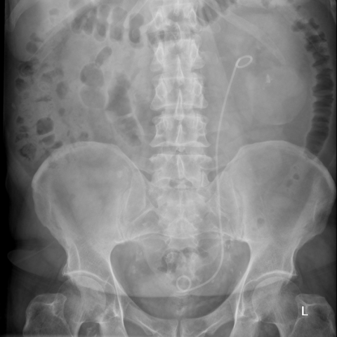
KUB X-RAY: Left JJ stent and left renal calculi.
Uroradiology & genital male imaging
Case TypeClinical Cases
AuthorsS.Chawla [1] J. C. Belfield [2]
73 years, male
[1] Kaude JV, Williams CM, Millner MR, Scott KN, Finlayson B. (1985) Renal morphology and function immediately after extracorporeal shock-wave lithotripsy. Amer J Roentgen 145(2):305-313.
[2] Alcover J, Rousaud A, Ruiz Marcellan Fj, Serrallach N, Serrate R. (1992) . Efectos adversos de las ondas de choque. Tema monográfico del LVII Congreso Nacional de Urología Ene Ediciones S.A. Cap. 3: Aspectos técnicos de la LEOC: 33. Cap 5: Efectos biológicos de las ondas de choque: 51-56.
[3] Collado Serra A, Huguet Pérez J, Monreal García de Vicuña F, Rousaud Barón A, Izquierdo de la Torre F, Vicente Rodríguez J. (1999) Renal hematoma as a complication of extracorporeal shock wave lithotripsy. Scand J Urol Nephrol 33(3):171-175
[4] Torrecilla Ortiz C, Matías López JJ, Contreras García J, Aguiló Luciá F, Camps Lloveras N, Riera Canals L. (1997) Renal hematoma after shockwave extracorporeal lithotripsy. Actas Urol Esp 21(8):752-757.
[5] Dhar NB, Thornton J, Karafa MT, Streem SB. (2004) A multivariate analysis of risk factors associated with subcapsular hematoma formation following electromagnetic shock wave lithotripsy. J Urol 172(6 Pt 1):2271-2274.
[6] Krishnamurthi V, Streem SB (1995) Long-term radiographic and functional outcome of extracorporeal shock wave lithotripsy induced perirenal hematomas. J Urol 154(5):1673-1675.
[7] Marini F, Martino F, Alfano V, Valente R. (1989) Computerized tomography and magnetic resonance imaging after ESWL. Arch Esp Urol 42(8):823-824.
[8] Tuteja AK, Pulliam JP, Lehman TH, Elzinga LW. (1997) Anuric renal failure from massive bilateral renal hematoma following extracorporeal shock wave lithotripsy. Urology 50(4):606-608.
[9] Hsin CY, Jung TS, Wen JW. (2008) Spontaneous bilateral subcapsular hematoma of the kidneys: a case report. J Taiwan Urol Assoc 19:228–31.
[10] Héctor Pastor Navarro, Pedro Carrión López, Jesús Martínez Ruiz, José Mª Pastor Guzmán, Mariano Martínez Martín, Julio A. Virseda Rodríguez (2009) Renal hematoma after extracorporeal shock-wave lithotripsy (ESWL). UROLÓGICAS ESPAÑOLAS 33(3):296-303
[11] Liang-Chung huang, Meng-Chia Chen, Che-Wei hsu (2010) Subcapsular Hematoma of Kidney with Rupture into the Perirenal Space After Extracorporeal Shock Wave Lithotripsy:. J Emerg Crit Care Med Vol. 21, No. 4
| URL: | https://eurorad.org/case/10180 |
| DOI: | 10.1594/EURORAD/CASE.10180 |
| ISSN: | 1563-4086 |
KUB X-RAY: Left JJ stent and left renal calculi.
Coronal CT KUB: Large left subcapsular renal haematoma
Axial CT KUB: Haematoma lying around the left kidney.
US: Echogenic mass around the left kidney
US:Echogenic mass around the left kidney
US: Left renal calculus
US: Normal Right Kidney


