Clinical History
An eleven week old female child presented with a left sided neck swelling which was first noticed shortly after birth. It was non tender, did not affect head control or movement and was not changing in size. There were no other relevent clinical findings. An ultrasound suggested the relevant diagnosis and was used to monitor progress.
Imaging Findings
An eleven week old female child was referred to the surgical team for assessment of a left sided neck swelling. The child’s mother had first noticed a palpable swelling several days after birth; there was no visible abnormality. Of note the child had had a forceps delivery but the marks from the forceps were at the level of the ears. The infant was in no apparent distress and had normal head movements and control. The swelling had not changed in size since first discovered.
On clinical examination, there was no visible abnormality. On mild neck extension, a firm non-mobile swelling was noted in the anterior body of sternocleidomastoid, measuring approximately 1.5cm. An ultrasound was obtained which showed a well defined solid mass in left sternocleidomastoid measuring 3 by 0.8 by 1.5cms (Figure 1a). The abnormality showed similar echogenicity to the underlying muscle. Several enlarged lymph nodes were also noted in the region. The normal right sternocleidomastoid was imaged for comparison (Figure 1b). The most likely diagnosis was felt to be fibromatosis colli, with other pathology such as rhabdomyosarcoma or metastatic lymphadenopathy being much less likely.
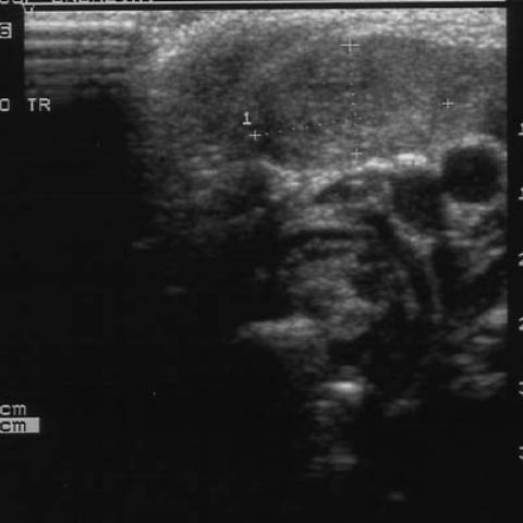
The infant was treated with passive physiotherapy of the neck, and initial follow up at two months showed the mass to have decreased in size both clinically and sonographically (Figure 2).
Within 5 months of presentation, the mass showed continued reduction in size (Figure 3). As the child was clinically improving, she was discharged to the care of her GP and no further imaging was performed.
Discussion
Fibromatosis colli (pseudotumour , sternocleidomastoid tumour of infancy) is a condition of unknown aetiology, affecting infants between the ages of two and four weeks. It is the most common cause of torticollis in this age group. It is a fibroblastic lesion, although whether neoplastic or reparative is uncertain. There may be a history of birth trauma, in up to 90% of cases. It is often associated with plagiocephaly and facial asymmetry. Clinically, it presents as a firm palpable swelling in the sternocleidomastoid, usually unilateral, in the lower one-third of the muscle and involving both sternal and clavicular heads. On ultrasound, an isoechoic mass is seen within the muscle. The lesion tends to grow rapidly, plateau and regress spontaneously, in 80-90% of patients, by the age of two years. The diagnosis is usually clear from clinical examination and ultrasound, although CT and MRI features are well described.
If biopsy is required in cases where the diagnosis is not clear-cut, fine needle aspirate (FNA) reveals mature fibroblasts in a granular background.
Management is with passive physiotherapy. In occasional cases, surgery may be required.
Differential Diagnosis List