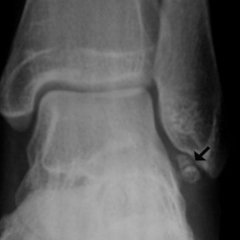
Figure 1
Ankle radiograph
Musculoskeletal system
Case TypeClinical Cases
AuthorsTheodorou SJ, Theodorou DJ, Barouchos N, Tsifetaki N.
21 years, female
[1] Vanhoenacker F, de Cuyper K, Williams H (2011) Normal anatomy and variants that simulate injury. In: Karantanas A (ed). Sports injuries in children and adolescents Berlin, Springer, p. 41-63
[2] Berg EE (1991) The symptomatic os subfibulare. Avulsion fracture of the fibula associated with recurrent instability of the ankle. J Bone Joint Surg 73A: 1251-1254 (PMID: 1890128)
[3] Ogden J, Lee J (1990) Accessory ossification patterns and injuries of the malleoli. Pediatr Orthop 10: 306-316 (PMID: 2113062)
[4] Powell H (1961) Extra centre of ossification for the medial malleolus in children: incidence and significance. J Bone Joint Surg 43B: 107-113
[5] Mellado J, Ramos A, Salvado E, Camins A, Danus M, Sauri A (2003) Accessory ossicles and sesamoid bones of the ankle and foot: imaging findings, clinical significance and differential diagnosis. Eur Radiol 13: 164-177 (PMID: 16440220)
[6] Hasegawa A, Kimura M, Tomizawa S, Shirakura K (1996) Separated ossicles of the lateral malleolus. Clin Orthop 330:157-165 (PMID: 8804286)
[7] Griffiths J, Menelaus M (1987) Symptomatic ossicles of the lateral malleolus in children. J Bone Joint Surg 69B: 317-319 (PMID: 3102500)
[8] Kono T, Ochi M, Takao M, Naito K, Uchio Y, Oae K (2002) Symptomatic os subfibulare caused by accessory ossification: a case report. Clin Orthop Rel Res 399; 197-200 (PMID: 12011709)
| URL: | https://eurorad.org/case/12186 |
| DOI: | 10.1594/EURORAD/CASE.12186 |
| ISSN: | 1563-4086 |
Coronal T1-weighted MR image
Coronal STIR MR image
Axial T2-weighted MR image