


Figure 1
Teaching Case
SectionAbdominal imaging
Case TypeClinical Cases
AuthorsTonolini Massimo, MD
Connected authors
27 years, male
Young immigrant from Ukraine suffering from high fever for one year, attributed to systemic lupus erythematosus and unsuccessfully treated with steroids in his native country. Physically found febrile, debilitated with generalized wasting, multiple cutaneous lesions and painful oral ulcers. Laboratory studies showed severe pancytopenia and increased C-reactive protein (200 mg/L).
Visceral leishmaniasis complicated by small bowel haemorrhage requiring transarterial embolisation
Myeloproliferative syndromes
Lymphoproliferative disorders / Lymphoma
Autoimmune syndromes e.g. systemic lupus erythematosus
Sepsis
Malaria
Typhoid fever
Tuberculosis
At admission, body CT (Fig. 1) revealed left lower lobe pneumonia and upper-normal-sized spleen with two unspecific focal lesions. Haemocultures and extensive serology (including HIV, Legionella, Pneumococcus, brucellosis and Widal reaction) tested negative. Visceral leishmaniasis was diagnosed on bone marrow with protozoa-laded histiocytes and confirmed by peripheral blood DNA polymerase-chain reaction.
A month following start of liposomial amphotericin-B therapy, the patient experienced haematemesis and melaena, initially treated by endoscopic clipping of gastric ulcers. After unsuccessful colonoscopy, persistent haemorrhage was identified by emergency CT (Fig. 2) as active intraluminal contrast extravasation in a jejunal loop, which was treated by transarterial embolisation. Persistent bleeding at CT (Fig. 3) required another embolisation session. Repeated CT (Fig. 4) confirmed stopped bleeding but detected a small bowel segment with thickened hypoenhancing walls suggesting post-embolisation ischaemia, confirmed at surgical exploration and resected.
Three weeks later, the patient was discharged in good clinical status with normalised blood count, C-reactive protein, and imaging appearances.
Leishmaniasis encompasses different disease forms resulting from vector-borne parasitic infection by protozoans classified as Leishmania species, and represents a major global health problem with an estimated prevalence of 12 million cases and over 350 million people at risk worldwide. Transmission occurs through bite of female haematophageous sandflies, including a zoonotic form (with dogs, rodents and other carnivores as reservoir) in the Mediterranean basin, Middle East, China and South America, and a human-human transmitted form in East Africa, India and Bangladesh. Also known as kala-azar, visceral leishmaniasis (VL) is the disseminated form caused by L. donovani and L. infantum that target the reticuloendothelial system macrophages, with a yearly incidence of 200-400,000 new cases mostly in children. Due to climate changes and increasing human migration, VL is gradually spreading from the impoverished endemic regions and is emerging as an opportunistic infection in HIV-positive individuals (particularly intravenous drug abusers with low CD4 T-cell counts) and is currently considered an AIDS-defining disease [1-4].
After a variable (weeks to years) incubation, clinical manifestations depend on the immune function and mostly include irregular fever, malaise, diarrhoea, abdominal pain and distension. Unless treated, VL causes progressive hepatosplenomegaly and pancytopenia from bone marrow suppression, and is eventually fatal in over 90% of cases from complications such as opportunistic superinfections and haemorrhages. Prognostic factors include HIV, advanced age, bleeding and jaundice. Diagnosis requires serology, identification of Leishmania in marrow microscopy, or polymerase chain reaction assay on peripheral blood and bone marrow aspirate [1-6]. Alone or in combination with other drugs, liposomal amphotericin-B is the preferred treatment, highly effective but toxic [7].
In conclusion, this often neglected tropical disease should enter the differential diagnosis of febrile syndromes with hepatosplenomegaly and/or haematologic abnormalities, particularly in people from endemic regions (such as India, Sudan and Ethiopia, Middle East and Brazil) and HIV-seropositive individuals. The most frequently involved gastrointestinal region is the duodenum, often with endoscopically normal mucosa [4, 5, 8]. Imaging features of VL are scarcely reported in literature and mostly include multiple nodular lesions in the spleen and liver [9, 10]. As this case exemplifies, in patients with suspected VL-related gastrointestinal bleeding multidetector CT-angiography is the preferred investigation technique, which provides accurate information about the presence or absence of intraluminal contrast extravasation representing active haemorrhage, and promptly allows correct choice of treatment including interventional embolisation or surgery [11].
References
[1] Abdalmaula GH, Barbadoro P, Marigliano A, et al. (2013) Human visceral leishmaniasis: a picture from Italy. J Infect Public Health 6:465-472 (PMID: 23999330)
[2] Alvar J, Velez ID, Bern C, et al. (2012) Leishmaniasis worldwide and global estimates of its incidence. PLoS One 7:e35671 (PMID: 22693548)
[3] van Griensven J, Diro E (2012) Visceral leishmaniasis. Infect Dis Clin North Am 26:309-322 (PMID: 22632641)
[4] Pintado V, Martin-Rabadan P, Rivera ML, et al. (2001) Visceral leishmaniasis in human immunodeficiency virus (HIV)-infected and non-HIV-infected patients. A comparative study. Medicine (Baltimore) 80:54-73 (PMID: 11204503)
[5] Buyukasik Y, Ileri NS, Haznedaroglu IC, et al. (1998) Fever, hepatosplenomegaly and pancytopenia in a patient living in the Mediterranean region. Postgrad Med J 74:237-239 (PMID: 9683980)
[6] McGwire BS, Satoskar AR (2014) Leishmaniasis: clinical syndromes and treatment. QJM 107:7-14 (PMID: 23744570)
[7] Balasegaram M, Ritmeijer K, Lima MA, et al. (2012) Liposomal amphotericin B as a treatment for human leishmaniasis. Expert Opin Emerg Drugs 17:493-510 (PMID: 23167833)
[8] Keramati MR, Khooei A, Aelami MH (2013) Visceral leishmaniasis with massive hematemesis and peripheral blood involvement. . Clin Lab 59:425-427 (PMID: 23724635)
[9] Bukte Y, Nazaroglu H, Mete A, et al. (2004) isceral leishmaniasis with multiple nodular lesions of the liver and spleen: CT and sonographic findings. Abdom Imaging 29:82-84. (PMID: 15160758)
[10] Raeymaeckers S, Docx M, Demeyere N (2012) MRI-findings of nodular lesions in an enlarged spleen, associated with visceral leishmaniasis. Eur J Radiol 81:2550-2553 (PMID: 22209428)
[11] Artigas JM, Marti M, Soto JA, et al. (2013) Multidetector CT angiography for acute gastrointestinal bleeding: technique and findings. Radiographics 33:1453-1470 (PMID: 24025935)
Case information
| URL: | https://eurorad.org/case/12830 |
| DOI: | 10.1594/EURORAD/CASE.12830 |
| ISSN: | 1563-4086 |
License
This work is licensed under a Creative Commons Attribution-NonCommercial-ShareAlike 4.0 International License.


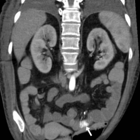


Figure 1

Tonolini M, Radiology Department, “Luigi Sacco\" University Hospital – Milan (Italy)

Tonolini M, Radiology Department, “Luigi Sacco\" University Hospital – Milan (Italy)

Tonolini M, Radiology Department, “Luigi Sacco\" University Hospital – Milan (Italy)

Tonolini M, Radiology Department, “Luigi Sacco\" University Hospital – Milan (Italy)

Tonolini M, Radiology Department, “Luigi Sacco\" University Hospital – Milan (Italy)
Figure 2

Tonolini M, Radiology Department, “Luigi Sacco\" University Hospital – Milan (Italy)

Tonolini M, Radiology Department, “Luigi Sacco\" University Hospital – Milan (Italy)

Tonolini M, Radiology Department, “Luigi Sacco\" University Hospital – Milan (Italy)

Tonolini M, Radiology Department, “Luigi Sacco\" University Hospital – Milan (Italy)

Tonolini M, Radiology Department, “Luigi Sacco\" University Hospital – Milan (Italy)

Tonolini M, Radiology Department, “Luigi Sacco\" University Hospital – Milan (Italy)
Figure 3

Tonolini M, Radiology Department, “Luigi Sacco\" University Hospital – Milan (Italy)

Tonolini M, Radiology Department, “Luigi Sacco\" University Hospital – Milan (Italy)

Tonolini M, Radiology Department, “Luigi Sacco\" University Hospital – Milan (Italy)

Tonolini M, Radiology Department, “Luigi Sacco\" University Hospital – Milan (Italy)

Tonolini M, Radiology Department, “Luigi Sacco\" University Hospital – Milan (Italy)

Tonolini M, Radiology Department, “Luigi Sacco\" University Hospital – Milan (Italy)

Tonolini M, Radiology Department, “Luigi Sacco\" University Hospital – Milan (Italy)
Figure 4

Tonolini M, Radiology Department, “Luigi Sacco\" University Hospital – Milan (Italy)

Tonolini M, Radiology Department, “Luigi Sacco\" University Hospital – Milan (Italy)

Tonolini M, Radiology Department, “Luigi Sacco\" University Hospital – Milan (Italy)

Tonolini M, Radiology Department, “Luigi Sacco\" University Hospital – Milan (Italy)

Tonolini M, Radiology Department, “Luigi Sacco\" University Hospital – Milan (Italy)
Figure 5

Tonolini M, Radiology Department, “Luigi Sacco\" University Hospital – Milan (Italy)

Tonolini M, Radiology Department, “Luigi Sacco\" University Hospital – Milan (Italy)

Tonolini M, Radiology Department, “Luigi Sacco\" University Hospital – Milan (Italy)

Tonolini M, Radiology Department, “Luigi Sacco\" University Hospital – Milan (Italy)