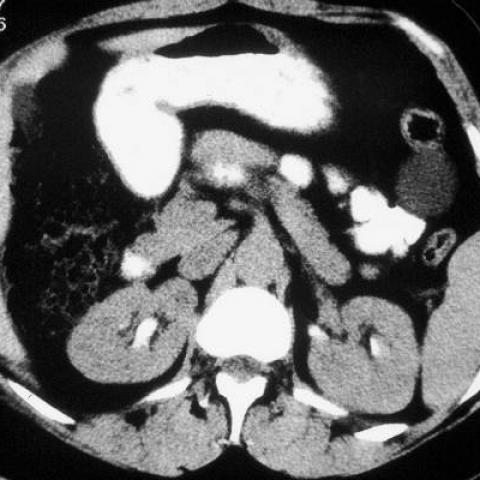
Mesothelial cyst at the splenic flexure


Abdominal imaging
Case TypeClinical Cases
AuthorsVinhais S, Monteiro M, Cunha TM
Patient44 years, female
Mesothelial cysts occur mainly in children and in young adults as asymptomatic abdominal masses. However, they can cause chronic abdominal pain or acute pain secondary to torsion, rupture, haemorrhage, infection or GI obstruction.
Macroscopically, they are thin-walled, unilocular cysts, usually with a serous content. Thus, on US mesothelial cysts appear as anechoic rounded lesions with acoustic enhancement. CT and MR reveal fluid-filled masses without a perceptible wall, presenting a low T1 signal and a high T2 signal in relation to their serous nature. Although these imaging findings are fairly characteristic, they are unspecific. Other types of mesenteric and omental cysts, such as lymphangioma, enteric duplication cyst, enteric cyst and nonpancreatic pseudocyst, may exhibit similar aspects. This is also the case with cystic neoplasms of peritoneal origin such as cystic mesothelioma, cystic teratoma and cystic spindle cell tumour.
The differential diagnosis of the large abdominopelvic lesion described in this case includes ovarian cyst, cystadenoma, cystic teratoma and paraovarian cyst. However, the simultaneous occurrence of two other lesions with identical appearance close to the viscera, points to a single diagnostic entity with peritoneal origin. Owing to their benign nature and loose attachment to the surrounding tissues, the treatment of choice is enucleation. Recurrence is extremely rare.
[1]
Ros PR, Olmsted WW, Moser RP, Dachman AH, Hjermstad BH, Sobin LH.
Mesenteric and omental cysts: histologic classification with imaging correlation.
Radiology. 1987 Aug;164(2):327-32. (PMID: 3299483)
[2]
Ravo B, Metwally N, Pai PB, Ger R.
Developmental retroperitoneal cysts of the pelvis. A review.
Dis Colon Rectum. 1987 Jul;30(7):559-65. (PMID: 3595378)
[3]
Stoupis C, Ros PP, Abbitt P, Burton S, Gauger J.
Bubbles in the belly: imaging of cystic mesenteric or omental masses.
Radiographics. 1994 Jul;14(4):729-37. (PMID: 7938764)
[4]
Vanek VW, Philips AK.
Retroperitoneal, mesenteric, and omental cysts.
Arch Surg. 1984 Jul;119(7):838-42. (PMID: 6732494)
| URL: | https://eurorad.org/case/1307 |
| DOI: | 10.1594/EURORAD/CASE.1307 |
| ISSN: | 1563-4086 |