Clinical History
A drug-addicted 26 years old homeless man presented to the emergency room with a left inguinal abscess refractory to antibiotic therapy and spiking fever of 39º. Physical examination revealed mixed bloody and purulent drainage. Leukocyte count was of 18.000. CT was requested in order to evaluate abscess size and extension.
Imaging Findings
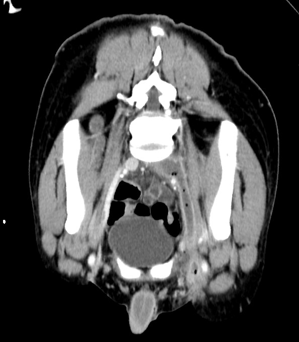
Emergency contrast-enhanced MDCT depicted a non-opacified left femoral vein extending to the external iliac, common iliac and inferior cava vein with prominent vessel wall enhancement and endoluminal gas bubbles indicating septic thrombophlebitis. Inflammatory changes were predominantly located at the site of the abscess. Multiples sites of injection were identifiable. Furthermore, subpleural lesions with central cavitations consistent with septic emboli were also present.
Discussion
Septic thrombophlebitis is an increasingly diagnosed and life-threatening event among intravenous drugs users, resulting from a non-sterile injection technique most usually in the femoral veins at the groin, when injection sites in the arms are no longer available [1-3]. Deep vein septic thrombophlebitis should be suspected, in the appropriate clinical setting, as a possible cause of persistent often spiking fever despite broad-spectrum antibiotic therapy. Leg oedema, flank or lower abdominal pain are frequent symptoms while a high leukocyte count and elevated inflammatory markers are common laboratory findings. Staphylococcus aureus is the most common causative organism [3, 5]. Contrast-enhanced multidetector CT with multiplanar reformations effectively detect the size and extent of deep septic thrombophlebitis. The affected veins are depicted enlarged with partially or entirely non-opacified lumen. An infected thrombus is suggested by intense vessel wall enhancement, endoluminal gas bubbles, and often by abnormal inflammatory hyperdense “stranding” of the surrounding fat planes [1-3]. Infections may spread to involve deeper soft tissues in the form of pyomyositis or even necrotizing fasciitis. MR imaging is the modality of choice for demonstrating the extent of these deeper soft-tissue infections. Subcutaneous and muscular abscess are also common injection site complications. Awareness should be placed in the fact that intravenous drug abuse is a multisystem disorder including vascular and infective complications affecting different organ systems, often synchronously. As in the case of our patient, pulmonary embolisation resulting from thrombus detachment and haematogenous dissemination is a frequent event. Lung involvement is showed as peripheral consolidations and ill-defined abscess-like nodules with frequent central cavitation [1, 4]. Recreational drug abuse is best regarded as a multisystem disease with potential complications involving many different organs, sometimes synchronously. It should be considered in any unexplained illness, especially in young people. Owing to the multisystem effects of drug abuse, the discovery of one complication should prompt the radiologist to search for signs of coexistent pathologic conditions, which may alter management. High clinical suspicion and prompt imaging assessment are therefore crucial for a timely diagnosis and treatment of this life-threatening condition and its further complications [1, 2].
Differential Diagnosis List
Femoral vein thrombophlebitis and septic pulmonary embolism from intravenous drug use
Aseptic venous thrombosis
Pseudo-aneurysm from arterial puncture
Final Diagnosis
Femoral vein thrombophlebitis and septic pulmonary embolism from intravenous drug use