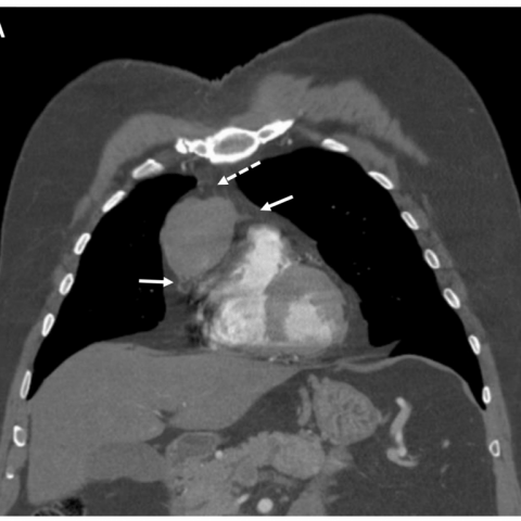
CASE 15651 Published on 09.04.2018

Unusual presentation of multiple endocrine neoplasia type 1: Primary thymic neuroendocrine tumour

Section

Chest imaging

Case Type

Clinical Cases

Authors

Haesung Lee BS1, Tarun Ramayya MD, Supriya Gupta MD, Darko Pucar MD, Nikhil Patel MD2, Jayanth H. Keshavamurthy MD3

(1) Senior Medical Student, Philadelphia College of Osteopathic Medicine
(2) MD-Assistant Professor, Department of Pathology, Augusta University
(3) MD-Assistant Professor, Department of Radiology, Augusta University.

1120 15th street,
BA-1411 30912 Augusta,
United States of America;
Email:
haesungle@pcom.edu
npatel4@augusta.edu
jkeshavamurthy@augusta.edu
Patient

60 years, male

Categories
Area of Interest Mediastinum ; Imaging Technique MR, Nuclear medicine conventional, Percutaneous, CT
Clinical History
60-year-old male patient with MEN-1 syndrome presented for evaluation of anterior mediastinal mass. Patient has had neuroendocrine tumour metastatic to the liver, status post modified Whipple procedure and exploratory laparotomy for partial hepatectomy. Patient also has history of parathyroidectomy x 2 and thyroidectomy x 2.
Imaging Findings
Computed tomography (CT) scan of the thorax confirmed a heterogeneous soft tissue mass with an unclear margin (dotted arrow) that is infiltrating the adjacent structures (white arrows) in the anterior mediastinum (Fig. 1 a and b). Magnetic resonance imaging (MRI) post contrast T1 imaging shows mildly heterogeneously enhancing anterio-mediastinal mass with irregular borders (Fig. 2). Radio-labelled fused Octreotide scan for MEN 1 evaluation showed a right anterior mediastinal mass that has increased radio-uptake (Fig. 3). Gross image of surgically removed mass shows greatest dimension 5.5 cm with additional dimensions 5.2 x 3.6 cm (Fig. 4 a). The tumour cells were arranged in wide trabeculae with irregular nests separated by thin fibrovascular stroma, and scattered abortive rosette-like structures. (Fig. 4 b). Tumour cells were diffusely positive for Synaptophysin (Fig. 4 c).
Discussion
A. Background:
Multiple endocrine neoplasia type 1 (MEN1) is an autosomal dominant syndrome that is associated with a large group of malignancies that are derived from embryonic neural crest tissue found in various organs such as parathyroid (90-97%), enterohepatic pancreatic tumours (30-80%), and anterior pituitary tumours (15-30%). [1]

B. Clinical Perspective:
Primary hyperparathyroidism and anterior pituitary tumours are surgically removed with good clinical outcomes. To date, enteropancreatic neuroendocrine tumours (NETs) are now the primary life-threatening manifestation of MEN1. [7] NETs are more aggressive, metastasise more often and have a poorer prognosis than other MEN 1 tumours. [4] Thymic NETs are the least common among the MEN 1 endocrine tumours with a prevalence of 2-8% in MEN 1 patients. [2] Additionally 25% of all thymic NETs occur in MEN 1 patients. Thymic NETs are also rare in the general population, accounting for 2-5% of all thymic tumours. [3] In a study of 80 cases of thymic NET, overall 5- and 10-year survival rates were 28 and 10 percent, respectively. [5] The poor prognosis may partially be due to late detection.

C. Imaging Perspective:
In all guidelines for MEN1 patients, NET localisation is an essential step in all aspects of their management. 111In-DTPA-octreotide (octreoscan) has been used for evaluation of pancreatic NETs. Recommended screening guidelines include chest X-ray and chest computed tomography (CT) every 3 years in male patient with MEN1 over the age of 25, but the benefits are still uncertain. [3] While CT imaging remains the recommended choice for initial cross-sectional imaging evaluation of mediastinal lesions, thoracic MRI provides definitive evaluation of mediastinal masses because of its superior tissue characterisation. [8] MR is the preferred modality for follow-up in patients with MEN 1 where repeated imaging may be required for prolonged surveillance due to the frequent recurrence of the disease. [8]

D. Outcome:
Surgical resection is the only curative technique in NETs. [8] Patient had video-assisted right thoracoscopic surgery for mediastinal mass resection with partial pericardial resection. Pathology report found this to be consistent with well-differentiated primary thymic NETs with invasion to neighbouring structures.

E. Teaching point:
Thymic NETs contribute to significant mortality in patients with MEN 1. Prophylactic thymectomy is considered in MEN1 male patients, but the benefits are uncertain as there are cases of recurrent thymic NET in patients after prophylactic thymectomy. [6] Thus, further studies of thymic NET cases are important to assess clinical management strategies.
Differential Diagnosis List
Thymic neuroendocrine tumour in multiple endocrine neoplasia type 1
Thymoma
Thymic neoplasm
Ectopic thyroid
Lymphoma
Teratoma
Final Diagnosis
Thymic neuroendocrine tumour in multiple endocrine neoplasia type 1
Case information
URL: https://eurorad.org/case/15651
DOI: 10.1594/EURORAD/CASE.15651
ISSN: 1563-4086
License