


Figure 1
Radiological features of Scurvy
SectionMusculoskeletal system
Case TypeClinical Cases
Authors
Samia Nesar, Hamza Abu-Jabeh, Oran Roche
Connected authors
12 years, male
Clinical History
A 12-year-old boy presented to the orthopaedic clinic with reduced mobility secondary to bilateral lower limb swelling and severe episodic knee pain. He was noted to have bleeding gums and his examination revealed ecchymosis of both lower limbs. He proceeded to have x-rays and MRI scans of both limbs.
Imaging Findings
MRI scan shows low signal regions on the T1-weighted images which are parallel to the endplates within the distal femoral and proximal tibial metaphysis, which are similarly seen in the proximal fibula [Fig. 2]. Matching areas of increased signal on STIR sequence through the tibial shaft and ankle joint metaphysis are seen[Fig. 3 + Fig. 4]. High STIR signal can also be noted in the distal femur intercondylar notch with low signal on T1-weighted sequences [Fig. 5 + Fig. 6]. There are subperiosteal collections overlying the posterior aspect of distal femurs bilaterally [Fig. 7 + Fig. 8]. Features of myositis within the tibialis anterior muscle and subcutaneous oedema with high signal throughout the medial and lateral gastrocnemius muscles are seen [Fig. 9]. The high STIR sequences are reduced post-treatment, showing radiological improvement [Fig. 10A -11B].
Discussion
Background
Scurvy is a clinical syndrome due to vitamin C deficiency (<11.4 µmol/L). Vitamin C is an essential co-factor required for the hydroxylation of proline and lysine and subsequent collagen synthesis. Its deficiency leads to the production of unstable pro-collagen chains predisposing to weakened bones and increased susceptibility to fractures.
In children, there are a number of risk factors associated with scurvy including malnutrition, malabsorption and neglect. Patients with neurodevelopment or psychiatric conditions are more likely to exhibit food selectivity [1,2]. Conditions that result in iron overload, such as thalassemia also deplete vitamin C causing scurvy [3].
Clinical Perspective
A thorough history including dietary habits and clinical suspicion is imperative to be able to diagnose scurvy. The early signs of scurvy are non-specific (fatigue, irritability and failure to thrive) with the classic symptoms of bleeding gums, ecchymosis, musculoskeletal pain and weakness presenting late. Severe prolonged deficiency can be life-threatening.
Imaging Perspective:
Radiological evidence of scurvy can be seen on plain radiographs of long bones, which are typically symmetrical and bilateral [4,5]:
- Cortical thinning (pencil-point cortex)
- Ground-glass osteoporosis
- Haemarthrosis and periosteal haemorrhages
- Scorbutic rosary: Angular knobbing of the costochondral joints
- Frankel line: Dense zone of provisional metaphyseal calcifications
- Trümmerfeld zone: Radiolucent line that is parallel to Frankel lines
- Wimberger ring: circular calcification surrounding the osteoporotic epiphyseal centre of ossification
- Pelkin spur: Calcification and periosteal elevation beyond the level of metaphysis
- Pelkin fracture: Metaphyseal avulsion fracture
- Corner angle sign: Irregular metaphyseal margins due to small infarctions
- Varus joint deformities and pathological fractures
MRI can detect early changes in scurvy. The commonest finding is diffuse multifocal decreased T1- weighted signal and increased T2-weighted signal within the bone marrow; with the metaphysis being the most affected. [6]
Outcome
The child was diagnosed with scurvy and was commenced on vitamin c replacement. His symptoms fully resolved. Overall, scurvy has an excellent prognosis.
Take-Home Message
Scurvy should be considered as a rare cause for limb and joint pain, particularly in high-risk children. It is important for radiologists to be aware of imaging findings found in scurvy and to appreciate the clinical context.
Differential Diagnosis List
Vitamin C deficiency (Scurvy)
Chronic recurrent multifocal osteomyelitis
Haematological malignancies (e.g. leukaemia)
Rickets
Lead poisoning
Non-accidental injury
Final Diagnosis
Vitamin C deficiency (Scurvy)
References
[1] Kothari P, Tate A, Adewumi A, Kinlin LM, Ritwik P. The risk for scurvy in children with neurodevelopmental disorders. Spec Care Dentist. 2020;40(3):251-9
[2] Nastro A, Rosenwasser N, Daniels SP, Magnani J, Endo Y, Hampton E, et al. Scurvy Due to Selective Diet in a Seemingly Healthy 4-Year-Old Boy. Pediatrics. 2019;144(3)
[3] Golriz F, Donnelly LF, Devaraj S, Krishnamurthy R. Modern American scurvy - experience with vitamin C deficiency at a large children's hospital. Pediatr Radiol. 2017;47(2):214-20
[4] Pan T, Hennrikus EF, Hennrikus WL. Modern Day Scurvy in Pediatric Orthopaedics: A Forgotten Illness. J Pediatr Orthop. 2021;41(3):e279-e84. 3
[5] Dr Pir Abdul Ahad Aziz Qureshi and Radswiki et al. Hypovitaminosis C. A Radiopaedia atricle.
[6] Gulko E, Collins LK, Murphy RC, Thornhill BA, Taragin BH. MRI findings in pediatric patients with scurvy. Skeletal Radiol. 2015;44(2):291-7
Case information
| URL: | https://eurorad.org/case/17536 |
| DOI: | 10.35100/eurorad/case.17536 |
| ISSN: | 1563-4086 |
License
This work is licensed under a Creative Commons Attribution-NonCommercial-ShareAlike 4.0 International License.

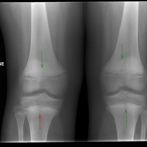

Figure 2

MRI of right knee, T1-weighted sequence, Coronal view
Figure 3

MRI of right knee, STIR sequence, Coronal view
Figure 4

MRI of left lower limb, STIR sequence, Coronal view
Figure 5

MRI of right knee, STIR sequence, Coronal view
Figure 6

MRI of right knee, T1-weighted sequence, Coronal view
Figure 7

MRI of right knee, STIR sequence, Coronal view
Figure 8

MRI of right knee, STIR sequence, Axial view
Figure 9

MRI of left lower limb, STIR sequence, Axial view


MRI of right knee, T1-weighted sequence, Coronal view
Luton and Dunstable Hospital, Luton, United Kingdom
Luton and Dunstable Hospital, Luton, United Kingdom

MRI of right knee, STIR sequence, Coronal view
Luton and Dunstable Hospital, Luton, United Kingdom
Luton and Dunstable Hospital, Luton, United Kingdom

MRI of left lower limb, STIR sequence, Coronal view
Luton and Dunstable Hospital, Luton, United Kingdom
Luton and Dunstable Hospital, Luton, United Kingdom

MRI of right knee, STIR sequence, Coronal view
Luton and Dunstable Hospital, Luton, United Kingdom
Luton and Dunstable Hospital, Luton, United Kingdom

MRI of right knee, T1-weighted sequence, Coronal view
Luton and Dunstable Hospital, Luton, United Kingdom
Luton and Dunstable Hospital, Luton, United Kingdom

MRI of right knee, STIR sequence, Coronal view
Luton and Dunstable Hospital, Luton, United Kingdom
Luton and Dunstable Hospital, Luton, United Kingdom

MRI of right knee, STIR sequence, Axial view
Luton and Dunstable Hospital, Luton, United Kingdom
Luton and Dunstable Hospital, Luton, United Kingdom

MRI of left lower limb, STIR sequence, Axial view
Luton and Dunstable Hospital, Luton, United Kingdom
Luton and Dunstable Hospital, Luton, United Kingdom


