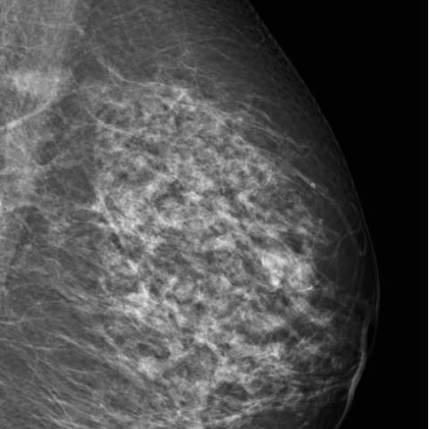
CASE 18097 Published on 05.04.2023

Granulomatous mastitis mimicking breast malignancy

Section

Breast imaging

Case Type

Clinical Cases

Authors

Penelope Moyle, James Tanner, Amit Agrawal

Cambridge Breast Unit, Cambridge University Hospitals, Cambridge, United Kingdom

Patient

60 years, female

Categories
Area of Interest Breast ; Imaging Technique Mammography, MR, Ultrasound
Clinical History

A 60-year-old, para 1 woman presented with a 3-week history of a painless right breast mass with no skin changes or other symptoms. She has a prior possible diagnosis of granulomatosis with polyangiitis although was not on treatment with no sequalae.

Imaging Findings

Mammography identified two right well-defined oval masses (Figure 1).

Ultrasound identified a 20mm mass containing small cystic spaces (Figure 2a) with no vascularity on Colour Doppler (Figure 2b). Ducts leading from the mass to the retroareolar and medial breast were expended by isoechoic soft tissue (Figures 2c, 2d).

Ultrasound-guided core biopsies were taken medially and laterally with clips placed at the sites of biopsies (Figure 2e).

Dynamic contrast MRI identified multiple rings enhancing masses, dilated ducts with enhancing walls extending to the retroareolar area, with type 1 kinetics (Figures 4a-d). The core biopsies identified abundant multinucleated giant cells, histiocytes, stellate cholesterol esters, periductal chronic inflammatory and eosinophilic proteinaceous acellular secretion. No malignancy, no necrotizing granulomas or vasculitis was identified. The appearances were consistent with granulomatous mastitis, B2.

As there were no symptoms apart from the initial mass, no treatment was initiated, and the breast masses clinically improved over 3 months.

Discussion

Idiopathic granulomatous mastitis (IGM) is a rare benign inflammatory breast disease characterised by lobulocentric granulomatous inflammation [1]. There are reported associations with pregnancy, lactation, oral contraceptives, hyperprolactinemia, autoimmunity and alpha-1 antitrypsin deficiency [2]. It is thought to be due to an initial insult to the ductal epithelial cells causing an inflammatory and then an idiopathic granulomatous response. Secondary granulomatous disorders must be excluded by histopathology, which include infections, autoimmune disease, sarcoidosis and diabetes [2].

IGM typically affects pre-menopausal, parous women with a history of lactation and presents as a unilateral palpable mass and can mimic infection with skin redness and skin thickening or mimic malignancy with a mass, peud’orange, nipple changes and less commonly lymphadenopathy [3].

Imaging can have a wide spectrum of appearances. On mammography it most commonly presents with asymmetry or masses. On ultrasound it can present with abscess formation, solid or cystic masses with or without ductal soft tissue extension, skin thickening and skin sinuses. MRI can demonstrate heterogeneously enhancing masses, rim-enhancing lesions, enhancing ducts and non-mass like enhancement. The T2 hyperintensity, and rim enhancement are presumed to represent microabscesses [4]. Imaging is important to guide core biopsy to obtain histopathology to exclude malignancy and other granulomatous inflammatory causes and to guide follow-up. Core-needle biopsy for cytopathologic examination and culture analysis are required to exclude malignancy and other benign inflammatory breast processes [5].

Patients with IGM have an excellent prognosis, although can have a relapsing-remitting cause before it burns out. Conservative treatment is the mainstay although oral steroids, immunosuppressive and prolactin-lowering medications can be given. Surgical excision may be an option for patients in whom medication therapy is unsuccessful.

This case is unusual as it presents in a post-menopausal woman, 20 years post pregnancy and lactation, with no other symptoms or signs except a mass, hence multifocal malignancy was initially suspected with ductal extension of tumour or in-situ disease. The avascularity of the main mass could have pointed to a more benign pathology, but tumours can be necrotic and have cystic components. The previous history of granulomas with polyangiitis could be considered although it is extremely rare [7] with no history of ongoing vasculitis nor necrotizing vasculitis in the pathology sample.  The MRI was typical of enhancing microabscesses and inflammatory ducts, which can occur in a variety of idiopathic or secondary granulomatous processes, but ductal carcinoma in situ can also demonstrate a ring-like enhancement pattern [8]. Taking representative core biopsies from different areas helps reduce sample errors. This case demonstrates the importance of clinical, radiological and pathology correlation as granulomatous mastitis can be a great mimic of benign and malignant disease processes.

Differential Diagnosis List
Granulomatous mastitis
In-situ and invasive breast carcinoma
Idiopathic Granulomatous Mastitis
Granulomatosis with polyangiitis
Infective mastitis
Specific granulomatous mastitis, secondary to systemic disease or infection
Final Diagnosis
Granulomatous mastitis
Case information
URL: https://eurorad.org/case/18097
DOI: 10.35100/eurorad/case.18097
ISSN: 1563-4086
License