

Uroradiology & genital male imaging
Case TypeClinical Cases
Authors
Muhammad Arif Saeed1, Shandana Khan2, Noman Khan3, Ayimen Khalid Khan3, Muhammad Nauman Shah4
Patient26 years, male
A 26-year-old male presented with mild left upper quadrant pain and discomfort that had started a month ago. The pain was dull, over the left lumbar region, and was unrelated to a specific time of the day, intensity with posture, diet or exercise. Patient had no other complaints and was otherwise in good health and had no comorbid conditions. There was no history of hematuria, dysuria, fever, urinary retention or urgency. Patient’s physical examination was normal and there was no tenderness over the lumbar region. The past medical and surgical history was unremarkable. The baseline laboratory investigations revealed no abnormality. Urine routine analysis was normal. The patient was suspected of having a renal calculus and CT KUB was obtained.
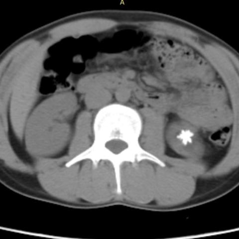
Computed Tomography of Kidneys, Ureters and Bladder (CT KUB) revealed a large star-shaped calculus with spiky projections in the lower pole calyx of the left kidney (Figure 1).
In addition, there was moderate hydronephrosis on the left side with mild cortical thinning in upper and lower pole regions (Figure 2). There was moderate dilatation of the left renal pelvis with abrupt calibre change at the pelvicureteric junction (PUJ), suggestive of partial pelviureteric junction obstruction. The left proximal ureter was normal in calibre. No calculus was seen in the right kidney, either ureter or the urinary bladder.
Surgery was offered to the patient, and he underwent an uneventful Anderson-Hynes pyeloplasty. Intraoperative findings included narrowing at the left PUJ. The redundant renal pelvis, PUJ and proximal ureter were resected, followed by ureteric anastomosis with the lower part of the renal pelvis. A J-J stent was placed. The calculus in the left lower pole was surgically removed (Figure 3).
The postoperative management course was uneventful, and the patient was discharged after 3 days. The DJ stent was removed during the patient's 1-week follow-up visit. Follow-up visit after one month was negative for any late complications. Patient was in a normal state of health without any active complaints.
Jackstone calculus is a unique type of urinary tract calculi which resembles toy jacks. It is composed of a dense central core of calcium oxalate dihydrate and radiating spicules from deposition of new minerals, giving a jackstone calculus its unique sea urchin appearance [1, 2]. Despite the scary appearance, jackstone calculi can be easily fragmented and usually require no surgical intervention [3]. In case reports, jackstone calculi in the bladder have been described; nevertheless, they are extremely rare in the upper urinary system. Here, we present a rare case of a solitary renal jackstone calculus along with a brief review of the literature.
The growth of jackstone calculus occurs at the tips of projections that contact the urinary tract wall, where abrasions cause the adherent mucoprotein to rub off, allowing calcium oxalate to accumulate [5, 12]. This growth pattern is what gives jackstone calculus its distinctive toy-jack appearance.
A pre-existing injury is thought to act as an inducer of calculus formation, which in our case was likely related to chronic low-grade obstruction at the pelviureteric junction. Jackstone calculus is often asymptomatic as the size and configuration restricts its distal passage; however, it can cause urothelial irritation, which manifests as hematuria [3]. Calculus may also cause discomfort aggravated by sudden movements and exercise.
Jackstone calculus was first described by Halsted in 1900 [4]. Everidge described jackstone calculus in 1927 in an 84-year-old man incidentally discovered during the course of suprapubic prostatectomy [5, 6]. A number of case reports have since been published describing jackstone calculi in the urinary bladder [7, 8]. Perlmutter et al. first described the sonographic appearance of jackstone calculus incidentally discovered in a 75-year-old male undergoing abdominal ultrasound [9]. The calculus was described as a star-shaped echogenic focus casting posterior acoustic shadowing. The calculus did not cause any symptoms and the patient did not follow up with a urologist.
In the literature, we found few case reports describing jackstone calculi in the upper urinary tract [8, 10, 11]. Goonewardena et al. described renal jackstone calculus in a 63-year-old woman, who had presented with intermittent painless hematuria [10]. The calculus in this case was fragmented with Holmium: Yttrium-Aluminum-Garnet [Ho: YAG] lasertripsy and the patient made an uneventful recovery. Lim et al. reported jackstone calculi in a 53-year-old male with a 1-month history of intermittent left flank pain, which was managed with percutaneous nephrolithotomy [11]. Similar to our case, this patient had PUJ obstruction visualized on CT and intraoperative pyelography. In our case, the PUJ obstruction was the primary reason for surgery, and the stone was removed to reduce the likelihood of a recurrence.
Teaching Points
Written informed patient consent for publication has been obtained.
[1] Singh KJ, Tiwari A, Goyal A (2011) Jackstone: A rare entity of vesical calculus. Indian J Urol 27:543–4. https://doi.org/10.4103/0970-1591.91449
[2] Brogna B, Flammia F, Flammia FC, Flammia U (2019) Jackstone Calculus: A Rare Subtype of Urinary Stone with a Sea-Urchin Appearance. Int J Nephrol Kidney Fail 4(5). https://doi.org/10.16966/2380-5498.166
[3] Sweeney AP, Dyer RB (2015) The “jackstone” appearance. Abdom Imaging 40:2906–7. https://doi.org/10.1007/s00261-015-0440-x
[4] Stewart JOR, O’connell ND (2008) Unusual vesical calculi. Br J Urol 33:215–8. https://doi.org/10.1111/j.1464-410x.1961.tb11607.x
[5] Brogna B, Flammia F, Flammia FC, Flammia U (2018) A Large Jackstone Calculus Incidentally Detected on CT Examination: A Case Report With Literature Review. World J Nephrol Urol 7:85–7. https://doi.org/10.14740/wjnu372
[6] Everidge J (1927) Jackstone Calculi. Proceedings of the Royal Society of Medicine 20:717–8. https://doi.org/10.1177/0035915727020005115
[7] Dyer RB, Chen MY, Zagoria RJ (2004) Classic signs in uroradiology. Radiographics 24(Suppl 1):S247–80. https://doi.org/10.1148/rg.24si045509
[8] Grases F, Costa-Bauza A, Prieto RM, Saus C, Servera A, García-Miralles R, et al (2011) Rare calcium oxalate monohydrate calculus attached to the wall of the renal pelvis. Int J Urol 18:323–5. https://doi.org/10.1111/j.1442-2042.2011.02741.x
[9] Perlmutter S, Hsu CT, Villa PA, Katz DS (2002) Sonography of a human jackstone calculus. J Ultrasound Med 21:1047–51. https://doi.org/10.7863/jum.2002.21.9.1047
[10] Goonewardena S, Jayarajah U, Kuruppu SN, Fernando MH (2021) Jackstone in the Kidney: An Unusual Calculus. Case Rep Urol 2021:1–3. https://doi.org/10.1155/2021/8816547
[11] Lim KY-Y, Sewell J, Harper M (2022) Jackstones in the renal pelvis: A rare calculus. Urol Case Rep 42:101994. https://doi.org/10.1016/j.eucr.2022.101994
[12] Heathcote JD, Rosen PA (2018) Jackstone Calculus. J Am Osteopath Assoc 118:627. https://doi.org/10.7556/jaoa.2018.140
| URL: | https://eurorad.org/case/18256 |
| DOI: | 10.35100/eurorad/case.18256 |
| ISSN: | 1563-4086 |
This work is licensed under a Creative Commons Attribution-NonCommercial-ShareAlike 4.0 International License.