Clinical History
This case deals with a newborn presented to our observation with clinical signs of bowel obstruction within 48 hours of birth, including abdominal distension and bilious vomiting.
Imaging Findings
A newborn presented to our observation with clinical signs of bowel obstruction within 48 hours of birth, including abdominal distension and bilious vomiting. There had been no passage of meconium per rectum. At physical examination a firm, rubbery mass was palpable in the lower quadrants of the abdomen. Other clinical and laboratory tests were unremarkable.
Plain abdominal film showed unevenly distended bowel loops with scarce air-fluid levels. At the level of the ileum a mass with a mottled appearance (ground glass-like) was detected, whereas the colonic lumen was free of gas.
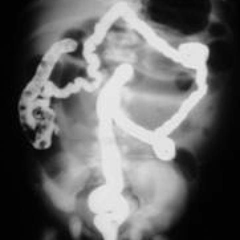
A contrast colon enema study, with a water-soluble contrast agent (Gastrografin), was performed showing a small unused colon; a continent ileo-cecal valve prevented reflux of contrast material in the terminal ileum. The clinical suspicion of meconium ileum was raised. The patient was operated on, because of the persistence of the occlusive state, and the pre-operative diagnosis was confirmed.
At laparotomy mid ileum bowel loops were distended containing thick and tenacious meconium. Bowel caliber got progressively smaller distally to the obstruction (distal ileum), with intestinal lumen impacted with multiple colorless pellets of inspissated mucus. A 12 F Foley balloon catheter was inserted and the small intestine was irrigated with multiple injection of a N-acetylcysteine solution (4 %). After few minutes, the sticky meconium and the mucous pellets moved into the colon relieving obstruction. The patient recovered without complications and the diagnosis of cystic fibrosis (CF), with a positivity to pilocarpine iontophoresis sweat test, was made.
Discussion
The genetically determined disease cystic fibrosis ( CF ) is the predominant cause of meconium ileus in infants. At the same time, meconium ileus is the earliest manifestation of CF and occurs in approximately 15% of CF patients. The meconium is extremely viscid and has an abnormal physico-chemical composition, such as less water content, decreased pancreatic enzymes, high concentration of undegraded serum proteins. The thick and viscous mucous secretion of the intestinal glands is the principal contributor to the sticky meconium causing bowel obstruction.
Roentgenographic studies are essential to confirm the clinical suspicion, establishing the diagnosis of meconium ileus and differentiating it from common conditions with similar presentation, including meconium plug syndrome, Hirschprung’s disease, left colon syndrome, small bowel atresia and functional immaturity of the intestine. There is no single pathognomonic sign that allows a radiographic diagnosis. Characteristic radiologic findings include unevenly distended bowel loops, a mottled mass in the lower tract of the small bowel, representing meconium that entraps small bubble of gas.
Colon enema is the most helpful diagnostic study and may be both diagnostic and therapeutic. It demonstrates a small, unused colon and may also outline the obstructing meconium mass in the distal ileum. The reflux of soluble hyperosmolar contrast material can relieve 60%-75% of uncomplicated meconium ileus, as a consequence of the fluid drawn into the bowel lumen and the mild mucosal irritation. If enemas are not successful, an operation is required. Prompt surgical intervention is also performed with major disease-related complications, such as volvulus, atresia and meconium peritonitis.
Differential Diagnosis List