Clinical History
The patient is a 19-year-old male who presented complaining of 3-week history of progressive headache unrelieved by analgesia and sudden onset of diplopia. On examination there was periorbital swelling and a left sixth nerve palsy. He was afebrile but his white cell count was 25. Social history included snorting cocaine.
Imaging Findings
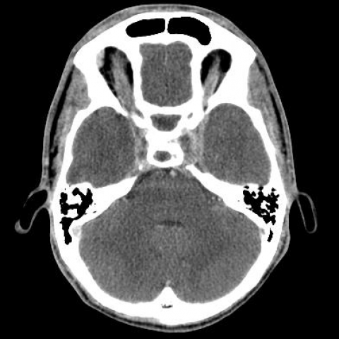
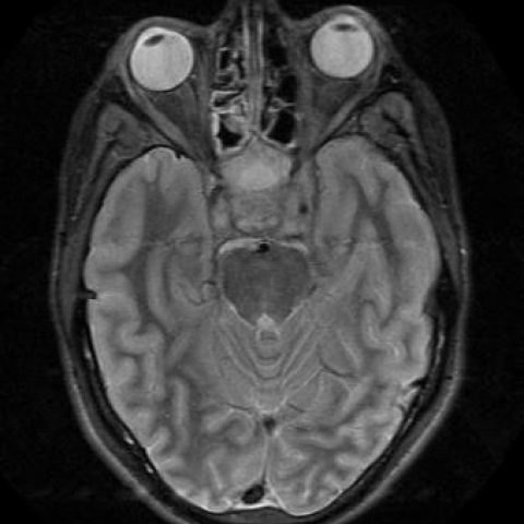
An unenhanced CT brain was requested from the emergency department which showed a swollen left cavernous sinus and mucosal thickening of the right frontal sinus, sphenoid sinus and ethmoidal air cells. A repeat scan post intravenous contrast administration was performed. This revealed multiple filling defects in both cavernous sinuses, enhancing left periorbital tissues and enhancing paranasal sinus mucosa suggestive of ongoing inflammation. The patient was started on intravenous Teicoplanin and Rocephine and IV heparin. The following morning an MRI scan confirmed the above findings. This showed proptosis on the left and bilateral swollen cavernous sinuses with intermediate signal intensity blood surrounding the internal carotid arteries. Absence of flow was also confirmed on MRV imaging. The patient was administered IV heparin and antibiotics for a total of two weeks after which he made a full recovery.
Discussion
The cavernous sinuses are made up of venous plexuses surrounded by a dural fold. The intracavernous portion of the internal carotid artery runs through the cavernous sinus surrounded by sympathetic nerve fibres. A number of cranial nerves also pass through, including the abducens nerve which lies lateral to the internal carotid artery, but medial to the oculomotor and trochlear nerves and the ophthalmic and maxillary divisions of the trigeminal nerve, which run superior to inferior within the lateral dural border of the cavernous sinus. [1]
The presenting symptoms of cavernous sinus thrombosis are usually encompassed into the cavernous sinus syndrome. This includes diplopia, Horner's syndrome and sensory loss of the first and second divisions of the trigeminal nerve with or without involvement of the pupil. [1]
Predisposing factors include brain and skull damage, intracranial and local regional infection, hormonal manipulation, post surgery, dehydration, connective tissue disease, malignancy, hypercoaguable states and idiopathic.
Both CT and MRI can be used for diagnosis. However, since most patients present as an emergency, CT scan is usually the initial investigation. Direct signs of cavernous sinus thrombosis include expansion of the cavernous sinus forming a lateral wall convexity rather than the normal concavity and multiple or single filling defects in enhanced scans. Indirect signs include dilatation of the superior ophthalmic vein, exophthalmos, soft tissue oedema and thrombi visualised in the veins and sinuses tributaries to the cavernous sinus. MRI is usually used to follow up after treatment, assess complications involving the pituitary gland and assess extension of infection to the adjacent meninges and brain. [2]
Immediate empiric antibiotic treatment should cover gram-positive, gram-negative and anaerobic bacteria. Treatment may then be narrowed, according to cultures and sensitivities. Surgical drainage of affected sinuses should be considered. Insufficient data exists regarding the value of anticoagualtion treatment as the entity is quite rare. The latter, used early, in conjunction with antibiotic treatment was found to significantly reduce later morbidity. Steroids are also used in cases of undrained abscesses. It does not improve mortality and morbidity but it reduces cranial nerve dysfunction. [3]
Differential Diagnosis List
Bilateral cavernous sinus thrombosis
Orbital cellulitis
Tolosa Hunt syndrome
Perneural spread of tumour
Final Diagnosis
Bilateral cavernous sinus thrombosis